|
| |
| Available exclusively from Henry Schein Ash Arcona |
 |
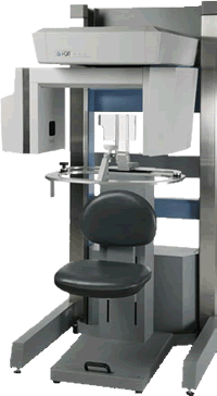
Why Cone Beam Technology?
Cone Beam 3-D images provide high-definition, three dimensional, digital data on precise anatomical information of all oral and maxillofacial structures.
• At reduced cost to the doctor
• With less radiation to the patient
|
 |
Why i-Cat?
The i-CAT enhances the way Dentists
see their patients. |
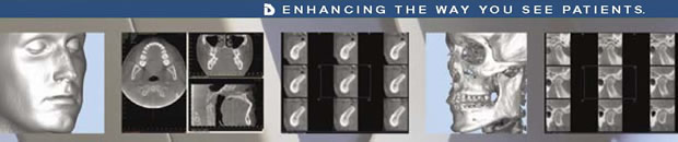
Surgical Predictability – The i-CAT gives dentists the complete anatomical information on bone and pathology prior to surgery resulting in less trauma and more predictable surgical outcomes.
Navigational Predictability – The anatomically accurate i-CAT images allow dentists to successfully create surgical plans and surgical guides prior to surgery.
Distortion Free Measurements –
The i-CAT produces anatomically accurate 3-D images used to create precise treatment plans leading to the most successful surgical procedures. |
Work Flow – The i-CAT takes 20-second open environment patient scans and, within minutes, the data is transferred to the workstation computer for the dentist to begin diagnosis and treatment planning.
Revenue Builder – Dentists are able to generate increased practice revenue through the billing process of i-CAT scans. The average charges across the country are $300–$400 per scan.
Case Acceptance – The i-CAT offers dentists a dramatic increase in case and treatment acceptance by allowing them to scientifically present information and diagnoses to patients. |
|
 |
For more information, contact
your HSAA Consultant
1-800-668-5558
or visit www.i-cat.com |
|
| Benefits
for the General Dentist |
Benefits
for the Orthodontist |
Benefits
for
Periodontists and
Oral Surgeons |
- Select the most suitable implant size and type.
- Achieve non-distorted and unmagnified measurements of the nerve canal height
- Provides the ability to find buccal/lingual width
- Provides complete 3-D information to optimize treatment planning and placement for implant surgery
- Offers ability to locate critical anatomy and determine if bone grafting or sinus lift is warranted
- Provides more accurate 3-D views of impacted molars, impacted cuspids, and other supernumerary anomalies
- 3-D views of critical structures for complete TMJ Analysis
- Assess airways and determine appropriate treatments
- Information can be utilized in implant planning software and to develop surgical guides further driving implant surgery to the general dentist
- Provides the most anatomically accurate images of the mouth, face, and jaw to determine proper diagnosis
|
i-CAT’s Optional Extended Field of View feature allows for a 22 cm. height scan, making it ideal for Cephalometric Reconstruction. Orthodontists are able to create a complete orthodontic work-up in one 20 or 40 second scan including:
Cephalometics SMV
Supernumerary Airway
Panoramic TM
Joints Impactions Spinal
- Enables location of impacted canines and supernumeraries
- Ability to check for root resorbtion
- In Class II or Overbite Patients you can determine the true reason for their mandible position
- Accurately measures problems to determine proper diagnosis
- Provide distortion-free 3-D views of critical anatomy surrounding the condyles
|
- Achieve non-distorted and unmagnified measurements of the nerve canal height
- Provides the ability to find buccal/lingual width
- Select the most suitable implant size and type
- Optimize locations and angulations for implant procedures
- Visualize impaction within the alveolar bone, location relative to adjacent teeth, and proximity to vital structures
- More accurate information resulting in less invasive surgery/decreased surgical time
- Diagnosis of bone morphology, joint space, and function for TMJ analysis
- High speed-scan for open jaw views
- Provides complete 3-D information to optimize treatment planning and placement
- Able to locate critical anatomy
- Determine if bone grafting or sinus lift
is warranted
- “Team Approach” with Restorative Dentists and other Specialists
|
 |
|